Optical Coherence Tomography (OCT)
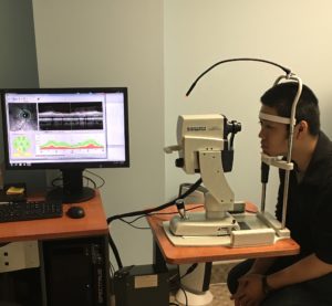
This month we are going to take a look at a piece of equipment called an Optical Coherence Tomographer (OCT) which was brought in to the office last May. This instrument is used on patients who have conditions that can affect the retina and optic nerve. The entire staff is pleased to bring this new piece of technology into the practice for our patients.
An OCT is a non-invasive test that takes images of the retina and optic nerve. While an OCT is similar to an ultrasound, these instruments use light to take cross section images of the retina; whereas an ultrasound uses sound to take the images. The images taken with an OCT allow the doctors to map out and monitor the thickness of each of the retinal layers. The images obtained during the test are used to monitor Age Related Macular Degeneration (AMD), Glaucoma, and other Diabetic Eye Diseases.
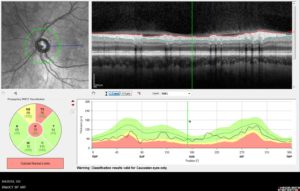
Furthermore OCT testing allows doctors to track and monitor changes to the fibers of the optic nerve for glaucoma patients . Therefore, patients with these conditions will return to the office several times in between their regularly scheduled eye exams to monitor the progression of their treatment plans. In addition, doctors can track and compare the results of progression and treatment over time as shown in the image below.
A question patients often ask is if an OCT can be performed without having to dilate their eyes. This is up to the doctor and patient to decide. A proper OCT image cannot be captured if the pupil is too constricted. Therefore, a dilation will allow the pupil to open more, allowing optimal light to get through for the images. However, if the doctor does decide to dilate the patient for the test, the dilation can cause light sensitivity and up close vision may be blurry for several hours after the exam.
Finally, OCT testing requires very little effort from the patient. The only thing patients need to do is look where the technician asks them to look and sit as still as possible. The technician and the instrument will do the rest of the work. This makes the testing quick and painless for the patient. To conclude the testing process, all of results are sent to the doctor for a final review. These final images will allow the doctor to determine the patients prognosis and continued treatment plan. Also, when the patient does return for their next follow up visit the doctor is able to compare the newest results to the previous visit. This will allow the doctors to see if the patients condition is progressing, allowing for a more accurate and proactive treatment plan.
For the doctors specific appointment hours please give our office a call. We will be happy to find an appointment time that fits into your busy schedules. Don’t forget to keep checking in on our webpage and our Facebook page for the most up-to-date information!
https://www.facebook.com/suburbaneyeinstitute
Dr. Laura Lukaszek: Monday – Thursday
Dr. Brian Mackey: Tuesday & Thursday – Saturday
Dr. Brenda Lynly: Monday & Wednesday